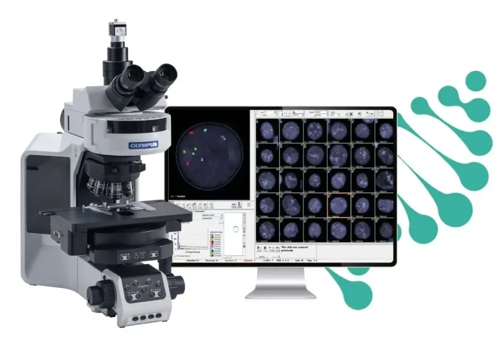
GenASIs HiFISH is a powerful tool for technologists working with FISH in pathology and cytogenetics laboratories. With this platform, laboratories can work faster, delivering more precise clinical results by using innovative tools and an optimal workflow.
Greater productivity for laboratories working with FISH
GenASIs FISH supports capture with a microscope manually or automatically, allowing any type of laboratory to considerably increase its efficiency. Automated features include:
Automatic height, focus and contrast adjustments.
Automatic XY coordinate sweep.
Automatic filter change.
Transformation and export of data to 3D.
BX63
For FISH tissue analysis, the GenASIs HiFISH platform offers technologists “Tissue Match”: a tool that allows analysis of the scanned location based on cell morphology, for tissues stained with H\&E or Immunohistochemistry.
GenASIs FISH Modules
GenASIs HiFISH is an image editing software that allows users to perform a multitude of enhancements, acquire images, and share them in a gallery. With GenASIs HiFISH, users can manually analyze each cell in FISH.
The GenASIs program “SpotScan” automatically analyzes cells in FISH. While FISHView performs analysis one cell at a time, SpotScan analyzes all the cells in all fields of view. Within seconds, SpotScan is enabled to provide statistical analyses of all counted cells, showing the number of signals and other clinical data while classifying them into categories.
Automatic segmentation based on morphological parameters and DAPI intensity.
User can define cell set based on signal count or amplification range.
Detects and enumerates small and weak signals.
FISHView
Manual System for FISH
In manual systems, FISHView assists in the analysis of slides viewed directly through fluorescence microscopes. The professional is responsible for manually adjusting the focus, exposure, and other parameters before capturing the images. After capture, FISHView offers tools to enhance the visualization and analysis of fluorescent signals.
Analysis Process
The operator manually captures images and uses FISHView to analyze the intensity and location of the signals. The interpretation of the results is performed by the professional, requiring experience in reading the images.
Advantages:
Full control over the capture and analysis process.
Flexibility to adjust specific parameters as needed.
Ideal for cases that require personalized and detailed analysis.
Limitations:
Slower process due to manual handling.
Susceptible to interpretation variations between different professionals.
Review Systems for FISH
In review systems, FISHView works with images automatically digitized by high-resolution scanners. The module applies algorithms to identify, quantify, and analyze fluorescent signals in an automated way, enabling faster and more accurate review.
Analysis Process
The slides are automatically scanned, and FISHView processes the images, highlighting the relevant signals. Then, the professional reviews the already analyzed results, being able to make adjustments or confirm the diagnosis.
Fast and accurate analysis with minimal manual intervention.
Greater consistency in results, reducing the possibility of human error.